The HPI Bioimaging Unit (HPI-BU) is one of the most modern and well-equipped Imaging Facilities in Greece and a member of the Greek Bioimaging-GR Consortium and the European Light Microscopy Initiative (ELMI- www.embl.org/elmi/). HPI-BU is an open-access platform aiming to provide advanced Light microscopy and image analysis services to HPI, national or international academic scientists.
The HPI-BU has created an international wide network with top imaging groups in the EU and the Institut Pasteur Paris that have largely contributed to the development and establishment of cutting-edge technologies in this Facility. European and National grants such as BIOIMAGING-GR – Ministry of Education Infrastructure Grant (2017-2020), Stavros Niarchos Foundation Grant: (2016-2020), KRHPIS I-II (2015-2018), NEUROSIGN EU FP7 Program (2010-2013) have supported the significant upgrading of the HPI-BU which has become a hub of a 2-photon Intravital Imaging system in the Bioimaging-GR Consortium, as well as fast live cell imaging systems.
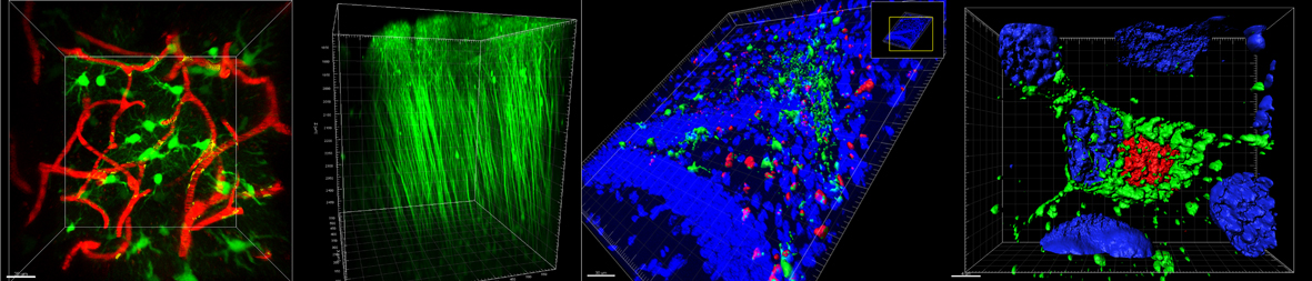
Τhe main state-of-the art methodologies currently in place in the HPI-BU are: a) 2- photon intravital imaging in the CNS of transgenic mice, b) short and long time live cell imaging and c) bioimage analysis using commercially available and open-access image analysis software.
The HPI-BU has emerged within the Institut Pasteur International Network as a node for advanced imaging training and scientific collaborations. The unit has organized many training activities and several publications have been published in peer review journals with the participation or acknowledgement of the HPI-BU.
Our strategic goal is to develop the appropriate tools and technology that will allow us to monitor in real time disease progression in a wide range of communicable and non-communicable disease models, to develop live cell Calcium imaging, as well as multiple parameter middle-throughput and high-throughput screening, aiming to the understanding of biological processes and screening for novel therapeutics.
Registration
New users of the Bioimaging Unit (BU) of HPI, as well as current users, who are starting a new project, should submit a project description in a form provided at the HPI-BU. We need all necessary information to organize on time introductory sessions for using the instruments, as well as for teaching and for support in sample preparation techniques and new imaging protocols.
External users
The HPI’s imaging systems can be used by external users, who should contact the responsible scientists of the Unit in order to arrange the exact time of the microscope booking.
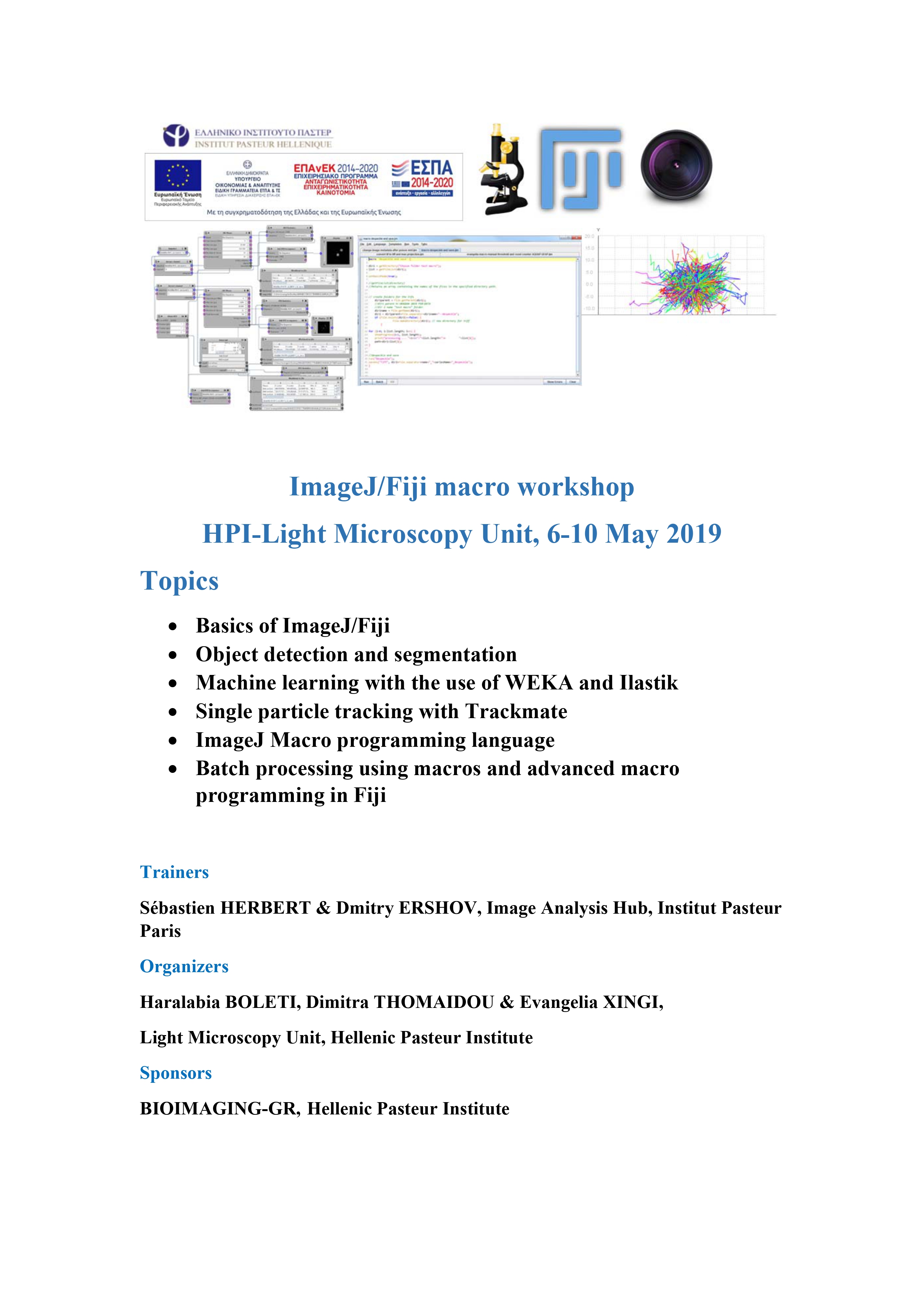
- ImageJ/Fiji macro workshop. 6-10 May 2019
A 5-day hands-on workshop was organized in the Hellenic Pasteur Institute in Athens Greece in collaboration with the Image Analysis hub of the Institut Pasteur Paris with the objective to provide theoretical and hands-on training in image analysis and automation tools.
The workshop programme included Lectures and hand-on sessions on: the basics of ImageJ/Fiji, object detection and segmentation, machine learning with the use of WEKA and Ilastik, single particle tracking with Trackmate, ImageJ Macro programming language, batch processing using macros and advanced macro programming in Fiji, while the last day of the workshop was dedicated to help the trainees analyze their own data and apply the new knowledge they gained.
The trainers were Dmitry Ershov and Sébastien Herbert, who kindly accepted our invitation to train the members of our Unit and external scientists on the ImageJ Macro programming language and machine-learning software. The trainees were 5 scientists from the Hellenic Pasteur institute (4 of which were members of the Bioimaging-GR program) and 3 external scientists from the University and other research centers in Athens.
Sponsors:
BIOIMAGING-GR, Hellenic Pasteur Institute
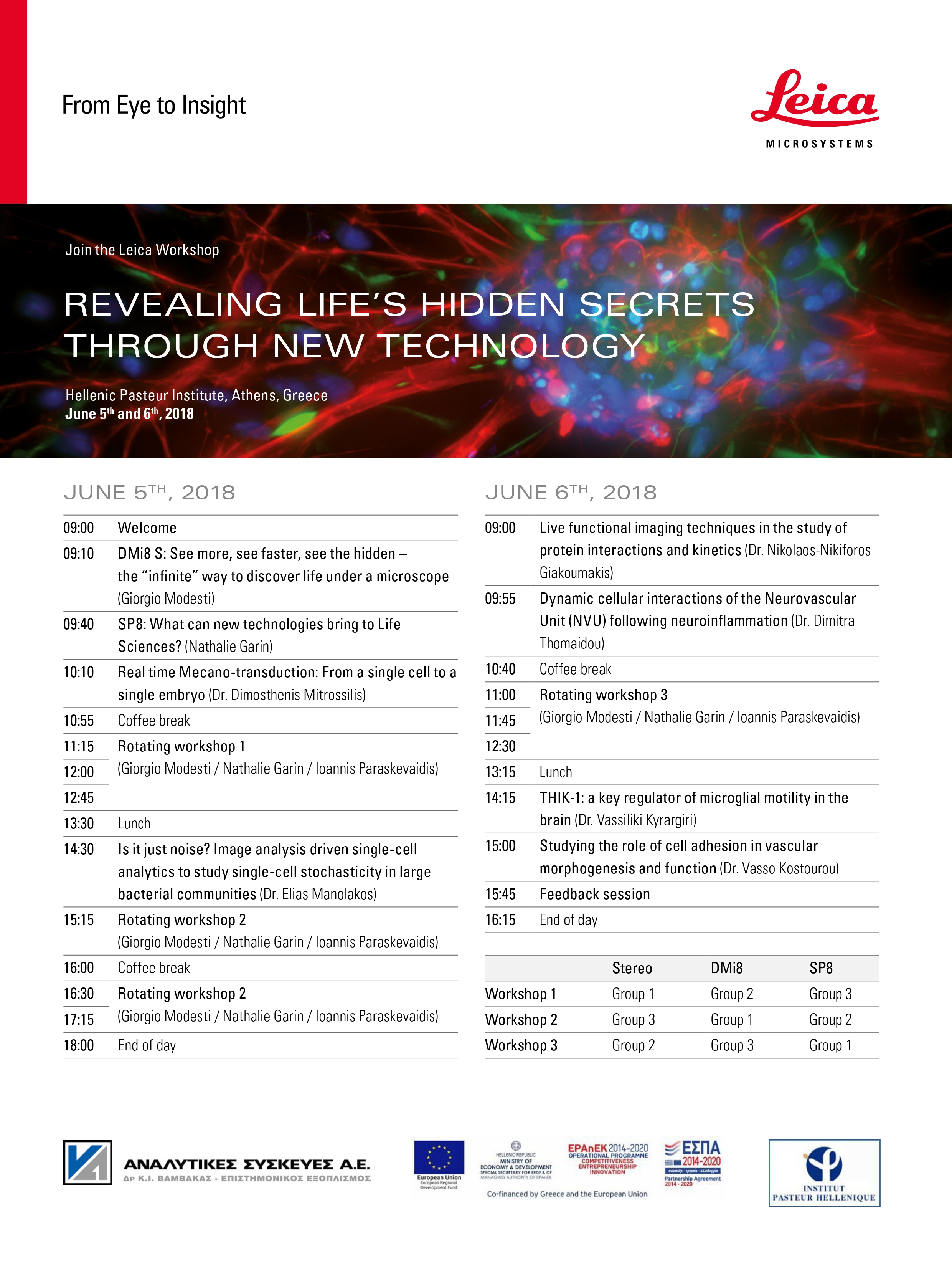
- «REVEALING LIFE’S HIDDEN SECRETS THROUGH NEW TECHNOLOGY» (5-6/06/2018) in collaboration with Leica Microsystems
The 2-day workshop was organized at the Hellenic Pasteur Institute in Athens, with the support of Leica Microsystems, the Bioimaging-GR program and the Hellenic Pasteur Institute. The objective of the workshop was to provide theoretical and practical training in advanced applications of microscopy in life sciences with an emphasis on 2-photon microscopy and confocal fluorescence microscopy through:
- Lectures by Leica Microsystems instructors and Greek scientists
- Practical training in the use of confocal microscopes
- Discussions with experts in the field of microscopy
35 trainees followed the theoretical and practical part, while a significant number of participants only attended the theoretical part of the seminar.
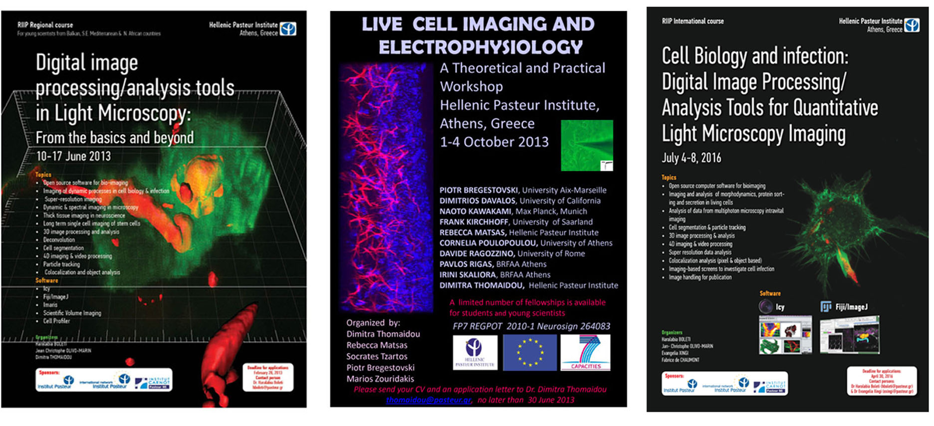
- RIIP International course “Cell biology and Infection: Digital Image processing/Analysis tools for quantitative light microscopy imaging” (4-8 July 2016)
A 5-day practical course was organized by the Hellenic Pasteur Institute in Athens Greece during 4-8 July 2016 with objective to provide theoretical and practical training in basic and advanced concepts and methods of Bioimage informatics. The course Program included:
- Lectures focused on concepts and methodology for Processing/Analysis of Light Microscopy Digital data (images and videos) and selected topics of Cell Biology and Biology of Pathogen Host interaction.
- Computer practical sessions on the quantitative analysis of microscopy image datasets using the open source software Icy and ImageJ/Fiji
- Informal discussions and tutorials with experts in the field
- European FP7 Regpot-2010 ‘Neurosign’ theoretical and practical workshop “Live Cell Imaging and Electrophysiology” (1-4/10/2013)
The workshop was conducted during 1-4 October 2013 in the Hellenic Pasteur Institute in Athens Greece. The aim of this workshop was to provide post graduate and post-doctoral scientists with a conceptual and practical understanding of CNS live cell imaging in whole animal level, as well as electrophysiology. During the practicals, the 20 selected participants had the chance to observe both live and video recorded experiments including surgical procedures for creating brain and spinal cord windows, intravital Multiphoton imaging and whole-cell and single-channel recordings, using the patch-clamp technique applied on mammalian cells. The funding was provided by the FP7 REGPOT 2010-1 EC Program “NeuroSign” that aims at the establishment of a Centre of Excellence for the study of Neurosignalling during nervous system function and dysfunction at the Hellenic Pasteur Institute.
- RIIP Regional Course “Digital image processing/ analysis tools in Light Microscopy: From the basics and beyond” (10-17/6/ 2013)
An 8-day course was organized in the Hellenic Pasteur Institute in Athens, Greece during 10-17 June 2013. The course objective was to provide theoretical and practical training in basic and advanced concepts and methods of digital image analysis and in the use of several open source and commercial software for quantitative assessment of fluorescence microscopy data through:
-
- Lectures
- Hands – on practical training sessions with participants’ microscopy data or data provided by the instructors.
- Informal discussions and tutorials with experts in the field.
- Practical sessions on the following image analysis software: Imaris, Scientific Volume Imaging (SVI), Image J/Fiji, Icy bioimage analysis, Cell profiler and Timm’s Tracking Tool.
|
The Scientists supporting the HPI-BU have participated either as members or as Scientific responsible persons in several grant applications succeeding to attract funding for the Facility either for organizing courses or for upgrading the equipment and development of new methodology from the following grants:
- International Network of Pasteur Institutes (IPIN) PTR Grant (2020-2022): “Microglia Imaging in Alzheimer’s Disease”. Scientific Responsibles: Dr. D. Thomaidou, Dr. M. Costa (Institut Pasteur Lille), Dr. J-Y. Tinevez, Institut Pasteur Paris.
- ΒΙΟΙΜAGING GR 2018-2020: “Hellenic Research Infrastructure for the Imaging and Observation of Fundamental Processes in Biology and Medicine” (MIS 5002755) which is implemented under the Action “Reinforcement of the Research and Innovation Infrastructure”, funded by the Operational Programme “Competitiveness, Entrepreneurship and Innovation” (NSRF 2014-2020) and co-financed by Greece and the European Union (European Regional Development Fund, coordinator Dr. Tavernarakis, IMBB Forth).
- Stavros Niarchos Foundations Sponsorships 2016-2020 “Development of innovative Biological products and health services for infectious and neurodegenerative diseases” (Coordinators Drs. R. Matsa and V. Myriagou)
- Ηellenic State Scholarship Foundation (Ι.Κ.Υ.) and German Burau for Academics’ Exchanges (DAAD): IKYDA program 2014-2015: Astroglia activation and reprogramming in brain injury and repair: analysis by in vivo imaging (2P-LSM) of the brain (Scientific Responsible Dr. D. Thomaidou).
- RIIP (Institut Pasteur International Network) International course 2016:
” Cell Biology and infection: Digital Image Processing/Analysis Tools for Quantitative Light Microscopy Imaging”. Scientific Responsible Dr. H. Boleti - FP7 EU program 264083 NEUROSIGN 2010-2013: Development of Centre of Excellence in Neurosciences (coordinators. Dr.S. Tzartos– Dr. R. Matsa & Dr. L. Probert (Dr. D. Thomaidou Responsible for WP of purchasing the Infrastructure equipment for in vivo imaging).
- RIIP (Institut Pasteur International Network) Regional course 2013:
“Digital image processing/analysis tools in Light Microscopy: From the basics and beyond”. Scientific Responsible Dr. H. Boleti - Competitive grant from the Greek General Secretariat of Research and Technology in the context οf European program “Human Networks for training in Research and Technology” 2004-2006 –Establishment of the Greek Light Microscopy Network Title: “Applications of Light Microscopy in Biomedical research and Diagnosis” In collaboration with the U. of Ioannina & U. of Crete Coordinator of the project and of the Network, HPI-LMU. (Scientific Responsible ¨Drs H. Boleti & D. Thomaidou)