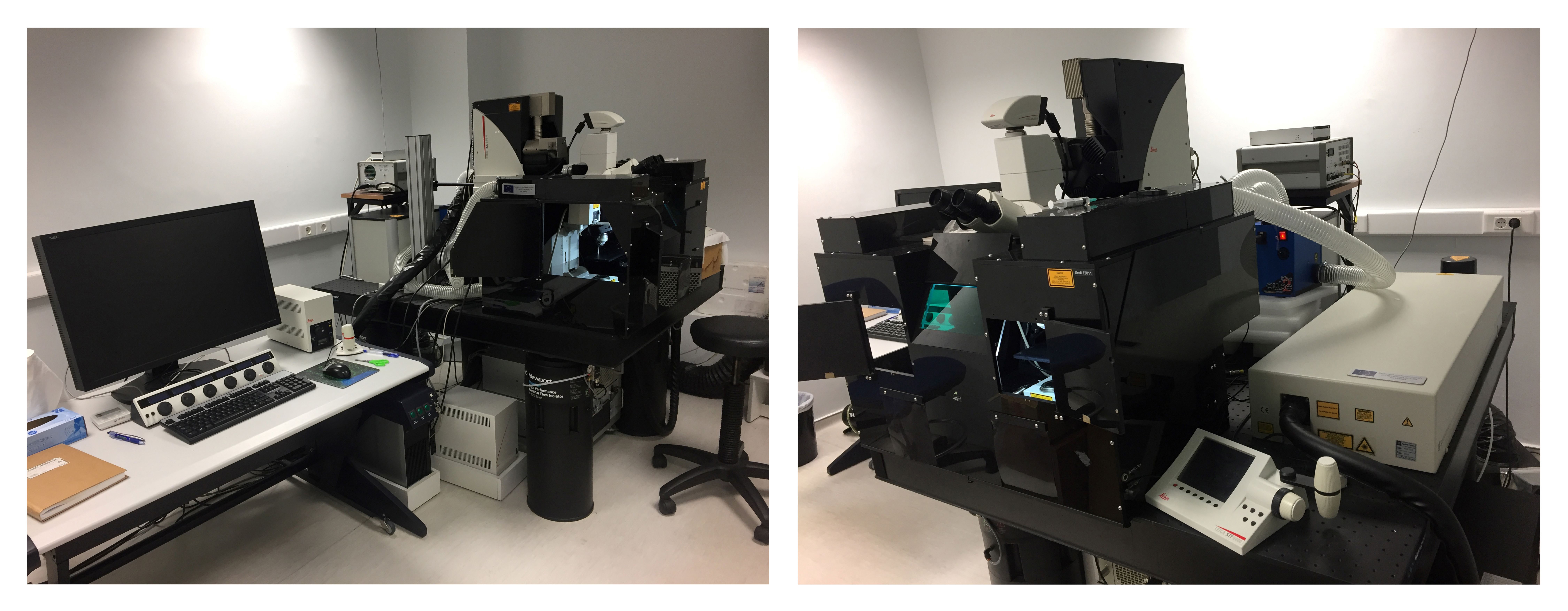
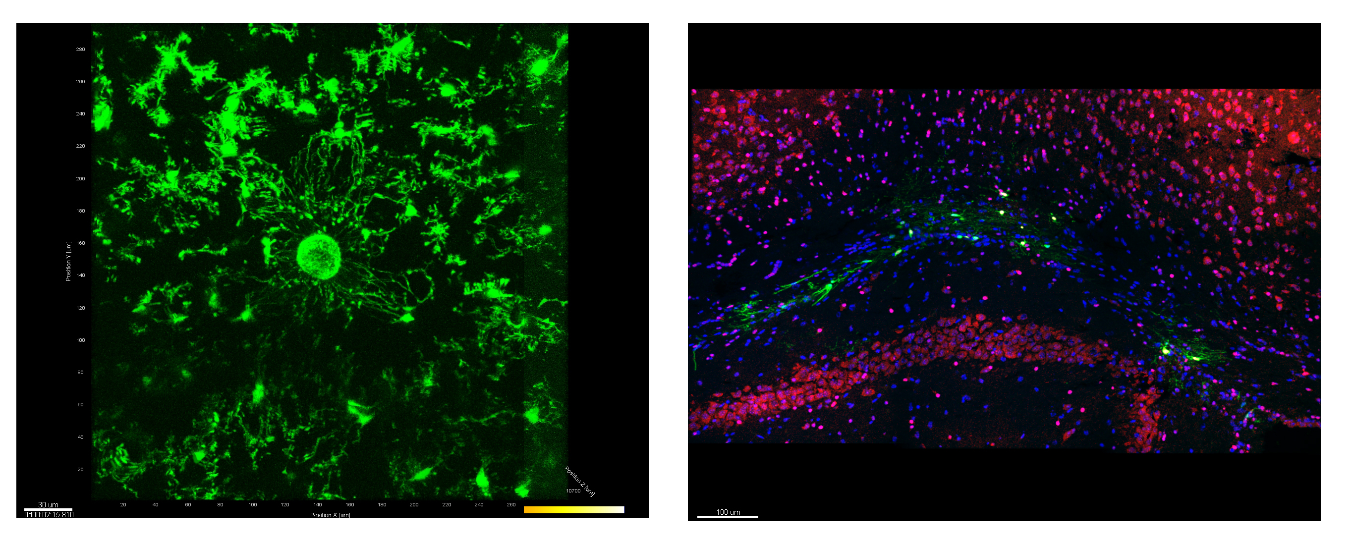
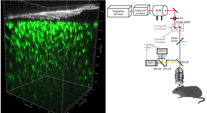
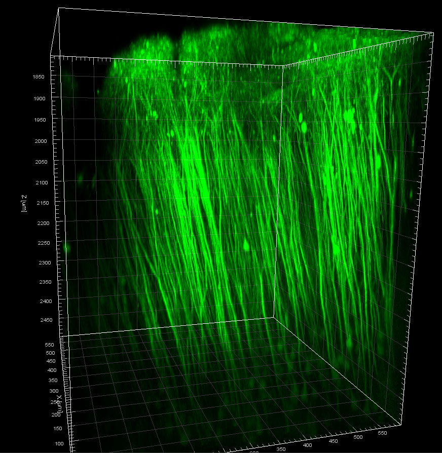
The Multiphoton Confocal system is appropriate for imaging of live biological samples in 3D and real time, as well as for imaging of small animals. The basic confocal system holds all the appropriate parts for Multiphoton microscopy applications (infrared (IR) laser source, multiphoton detectors, multiphoton fluorescence separating filters). More specifically, the technical characteristics of the system are:
A. Automated fixed-stage up-right confocal microscope, with alterations for also performing multiphoton microscopy bearing:
- Automated scanning stage, for acquisition of optical sections in Z-axis (z-galvo for precise movements, z-wide for coarse movements)
- Automated and coded disc for 6 objective lenses
- Head equipped with flat ocular lenses 10X
- Detector of transmitted light that allows simultaneous projection of fluorescence and transmitted light images.
- Halogen light source 12V, 100W for transmitted light applications.
- External Hg light source 120W for fluorescence applications, equipped with diaphragms for volume fluctuations and optical fibers for light transfer.
- Disc for holding 5 fluorescence filter sets and 3 filter sets for the UV (340/425), blue (450/490) and green (515/561) wavelengths.
- Flat apochromatic lenses of excellent quality, corrected to infinite and to UV. Dry lenses: 10X (NA 0.3) and 20X (NA 0.7), Oil-immersion lenses: 40X (NA 1.3) and 63X (NA 1.4).
- The microscope body is surrounded by dark environmental chamber (with controlled temperature, humidity and CO2 concentration) for performing live cell imaging experiments.
- The entire system is placed on a special anti-vibration table.

B. The confocal scanning system contains:
- High analysis scanning head for scanning at: a) xyz-axis, b) xzy-axis, c) temporal image scanning at xt, xyt, and xyzt, d) zoom between 1x and 64X and e) spectral scanning xλ, xyλ and xzλ, Scan resolution: Up to 8192 x 8192 pixels, Scanning speed: upto 1400 Hz
- 4 laser sources (405Diode, Argon, DPSS 561, HeNe 633) and laser lines covering wavelengths from UV to IP (405-650nm), more specifically at 405, 458, 476, 488, 496, 514, 561 and 633nm.
- Acousto-optic tunable filters (AOTF) for selecting specific wavelengths for excitation light
- Two photo-multipliers (PMTs) for confocal detection, a HyD3 detector of enhanced sensitivity and quantosome yield, and a PMT trans detector for phase-contrast detection. All these detectors have independent gain, offset and spectral position adjustments.
- The system’s software LAS AF allows the full control of the confocal microscope, laser sources and scanning head, image acquisition and storage, 3D image reconstruction from serial optic sections, creation of time- lapse movies and image processing.
- The system also allows FRET and FRAP applications, as well as pre-programmed workflow applications (FRET and FRAP wizard) and contains optic system for laser enhancement for FRAP application, that can be selected and controlled through the software (FRAP booster).

C. For Multiphoton applications in thick samples and whole small animals the system contains:
- Spectra Physics Mai Tai DeepSee infrared laser source
- Laser intensity controllers: a continuously adjustable electro-optical modulator (EOM) and a mechanical polarizer (half-wave plate)
- Carrier of 2 external non-descanned detectors (NDDs) for maximum signal collection. The available filter configurations are: FITC/TRITC, DAPI/TRITC, DAPI/FITC and CFP/YFP.
- One ceramic water-immersion objective lense 25X with high NA of 0.9 and working distance of 2500μm, dedicated for the transmission of the IR laser and appropriate for whole animal imaging.
- Multi-axis mouse holder stage with 4 incorporated drives for manual movement in the x, y dimensions and rotation mounts with graduated scale for repetitive brain imaging (provided by Prof F. Kirchhoff laboratory).


